Scripps Research scientists honored with prestigious CZI awards to address diseases tied to aging

Via The Scripps Research Institute News and Events Special Feature:

Christopher Parker and Giordano Lippi are unraveling the metabolic and protein mechanisms, respectively, that go awry in aging and disease—including Alzheimer’s.     

February 29, 2024

LA JOLLA, CA—How do our cellular mechanics change as we age, including in diseases like Alzheimer’s? Two Scripps Research scientists have been granted prestigious awards from the Chan Zuckerberg Initiative (CZI) to help answer that expansive question.

Christopher Parker, PhD, associate professor of chemistry, is a co-recipient of an Exploratory Cell Networks grant to chart the RNA metabolic networks that contribute to aging and disease. At the same time, Giordano Lippi, PhD, associate professor of neuroscience, has been granted the Collaborative Pairs Pilot Project Award to determine how a type of non-coding RNA impacts neurodegeneration. Both projects will reveal critical insights about the mechanisms that differentiate health from disease. 

Shedding light on RNA metabolism

Dysregulation of RNA processing can lead to altered gene regulation and disrupted cellular responses, but there is much about this process that remains unknown. The Exploratory Cell Networks grant will provide $1.25 million in funding to Parker’s lab over the course of three years, supporting the creation of strategies to investigate dynamic regulation RNA interactions, processing and metabolism. Parker will work alongside other grantees at neighboring institutions, including Christian Metallo, PhD, professor in the Molecular and Cell Biology Laboratory and Daniel and Martina Lewis Chair at the Salk Institute for Biological Studies; and Eugene Yeo, PhD, professor of Cellular and Molecular Medicine, and Nigel Goldenfeld, PhD, the Chancellor’s Distinguished Professor of Physics, at the University of California, San Diego. Together, they will use these technologies to clarify how cells respond and adapt to genetic and environmental stresses. This includes aging-related diseases linked to RNA dysregulation, such as Alzheimer’s disease.

“I am honored to receive this grant from the Chan Zuckerberg Initiative, which will empower our collaborative efforts to unravel the complex interplay between RNA and associated protein regulators,” Parker says. “Working hand-in-hand with other esteemed institutions across San Diego, we aspire to uncover new insights that could shape our understanding and treatment of different diseases, particularly those influenced by aging.” 

Collaboration is a central component of the Exploratory Cell Networks grants, with each award comprising researchers from at least three different institutions. By building these regional networks of investigators, CZI intends to unite technology development and accelerate the grant’s overarching goal: to better understand health and disease.

“We believe that scientific collaborations bring together new ideas and approaches that rapidly accelerate the pace of progress, making it natural for us to foster these enabling, cross-lab partnerships,” said Scott Fraser, CZI Vice President of Science Grant Programs. “Collaboration and building communities of researchers are central to our grantmaking strategy, and we’re excited to see what our new Exploratory Cell Networks teams will accomplish.”

At Scripps Research, Parker’s lab develops new tools and technologies to discover how proteins function in every human cell type, with the intent to develop effective therapeutics for a wide range of human diseases. Recently, he published a Nature Chemical Biology study highlighting a new technology that uncovers the ‘druggable’ sites on proteins for any human disease. In a separate Nature Chemical Biology study, Parker and his colleagues developed a small molecule that blocks the activity of a protein linked to autoimmune diseases, including lupus and Crohn’s disease.

A new approach to treating neurodegeneration

While aging often correlates with neurodegenerative diseases, how the aging brain becomes susceptible to these conditions is still unknown. With the Collaborative Pairs Pilot Project Award, Lippi will work with Eugenio Fornasiero, PhD, group leader in the Department of Neuro- and Sensory Physiology at the University Medical Center Göttingen, to understand the role and therapeutic potential of a type of RNA called microRNA (miRNA) in aging and neurodegeneration. Lippi and Fornasiero will receive $200,000 over 18 months, while also receiving support, mentoring and collaborative interactions of CZI’s Neurodegeneration Challenge Network.

CZI developed the Collaborative Pairs Pilot Project Awards to catalyze new collaborations and scientific partnerships, while springboarding early-stage projects they recognize as “bold, creative and ‘out-of-the-box.’”

For Lippi and Fornasiero’s project, they will be investigating how miRNA networks impact protein dynamics in the context of aging-related diseases. miRNAs are small, single-stranded molecules of RNA that, unlike their mRNA counterparts, do not code for proteins. Instead, they regulate which genes are turned on and off in the cell. Lippi and Fornasiero will explore how these miRNA networks affect protein production and degradation—particularly what goes awry during the aging process and corresponding diseases.

In previous research, scientists have found that proteins associated with neurodegenerative diseases aren’t replaced as efficiently, causing the brain to accumulate molecular damage. Lippi and Fornasiero are striving to identify the specific miRNA nodes that can prevent—or even reverse neurodegeneration—when targeted. To do this, they will use a new model that precisely maps miRNA-target interactions across different brain cells.

“I am deeply grateful for CZI’s support, as it fuels our pursuit to unravel the complex role of miRNA in shaping protein dynamics, while holding promise for transformative insights into neurodegenerative diseases,” says Lippi. “We aim to illuminate new paths of potentially treating these devastating diseases, which impact millions of people around the world.”

About Scripps Research

Scripps Research is an independent, nonprofit biomedical institute ranked one of the most influential in the world for its impact on innovation by Nature Index. We are advancing human health through profound discoveries that address pressing medical concerns around the globe. Our drug discovery and development division, Calibr, works hand-in-hand with scientists across disciplines to bring new medicines to patients as quickly and efficiently as possible, while teams at Scripps Research Translational Institute harness genomics, digital medicine and cutting-edge informatics to understand individual health and render more effective healthcare. Scripps Research also trains the next generation of leading scientists at our Skaggs Graduate School, consistently named among the top 10 US programs for chemistry and biological sciences. Learn more at www.scripps.edu

About the Chan Zuckerberg Initiative

The Chan Zuckerberg Initiative was founded in 2015 to help solve some of society’s toughest challenges — from eradicating disease and improving education to addressing the needs of our local communities. Our mission is to build a more inclusive, just, and healthy future for everyone. For more information, please visit chanzuckerberg.com.

Scripps Research awarded NIH BRAIN Initiative grant to map undiscovered parts of the central nervous system

Via The Scripps Research Institute News and Events Special Feature:

The $2.3 million award will support development of the first molecular toolkit that uncovers the brain’s “translatome” to better understand neural cognition, behavior and disease.

August 22, 2022

LA JOLLA, CA—Across science and medicine, the brain remains one of the most mysterious and complex parts of the human body. Scripps Research associate professor Giordano Lippi, PhD, has been awarded a grant from the National Institutes of Health (NIH) Brain Research Through Advancing Innovative Neurotechnologies® (BRAIN) Initiative to develop a new technology that reveals a major neural component and enhances our understanding of human brain function.

The highly regarded grant will provide $2.3 million in funding over one year, split evenly through a collaboration between Lippi’s lab and the Gene Yeo lab at the University of San Diego, California (UC San Diego). This funding will help advance a technology system that maps the brain’s “translatome”—the full collection of mRNAs (the genetic material that carries protein-coding information) that are highly translated into proteins (the effectors of all functions in the cell).

“So much of the human brain is still a puzzle to researchers today, and we need enhanced tools to better understand the molecular mechanisms underlying everything from cognition to behavior to disease,” says Lippi, who is an associate professor both in the Department of Neuroscience and Dorris Neuroscience Center, as well as an adjunct professor in the Department of Neurosciences at UC San Diego. “I am honored to receive this grant and help answer some of neuroscience’s biggest questions alongside a team of leading RNA experts, computational biologists and molecular neuroscientists.”  

Lippi and Yeo’s technology toolkit is known as Ribo-STAMP (Surveying Targets by Antibody-free Mutation Profiling), which measures—at single-cell level—how frequently a ribosome (the protein synthesis machine) binds to mRNAs. This an excellent proxy of protein translation, the final step of protein synthesis. Current related technologies like single-cell RNA sequencing rely on measuring only mRNA levels, which is a poor predictor of protein levels. In other words, Ribo-STAMP shows the full translational picture that is a much better representation of gene expression patterns in the brain.

Ribo-STAMP has the potential to bring this critical aspect of gene expression to light for the first time and help scientists to better understand—and even predict—how the translatome impacts the neurobiological processes underlying physiology, plasticity and disease. To achieve this, the team, supported by the grant, will expand Ribo-STAMP to be used for different neuroscience applications and to map the translatomes of specific neural cell types.

“With this funding, we can fully develop the most informative and scalable molecular profiling technology, and as a result, finally uncover the brain’s translatome at high resolution,” Lippi adds.

Whitehall Foundation honors two pioneering Scripps Research scientists

Via The Scripps Research Institute News and Events Special Feature:

LA JOLLA, CA – Neuroscientists Giordano Lippi, PhD, and Li Ye, PhD, of Scripps Research have received grants from the Whitehall Foundation to support their studies into brain function. Each researcher will receive $225,000 over three years.

“The Whitehall Foundation has a history of supporting young neuroscientists at the beginning of their tenure-track careers,” says Lippi, an assistant professor at Scripps Research and member of the Dorris Neuroscience Center. “Many awardees have gone on to very successful careers. It is therefore a true honor to receive this award.”

“It's a great honor to be acknowledged with this prestigious new faculty award in neuroscience,” says Ye, an assistant professor at Scripps Research and member of the Dorris Neuroscience Center. “Having this award will allow us to try out-of-the-box, high-reward projects.”

In the Lippi lab, the new funding will support research into microRNAs, the regulatory molecules that control brain development. The researchers plan to investigate a microRNA molecule called miR-218, which has been linked to epilepsy and cognitive impairments in human patients. Lippi says this work will help scientists unravel the molecular mechanisms underpinning healthy brain development.

“This project in particular, like most of the work in our lab, is at the intersection of circuit neuroscience and hard-core molecular biology,” says Lippi. “The Dorris Neuroscience Center is the perfect place for this kind of research, because of the expertise of my colleagues and the highly collaborative environment.”

The separate award to the Ye lab will support research to identify the brain circuits that drive organisms to eat more food when they are in a cold environment. One question researchers want to answer is how acute energy consumption affects the control of appetite.

“Scripps Research has a long history of promoting interdisciplinary research,” says Ye. “This project is a great example of cross-field research involving both neuroscience and metabolism.”

The Whitehall Foundation is a not-for-profit corporation founded in 1937 to support basic neurobiology research in the United States.

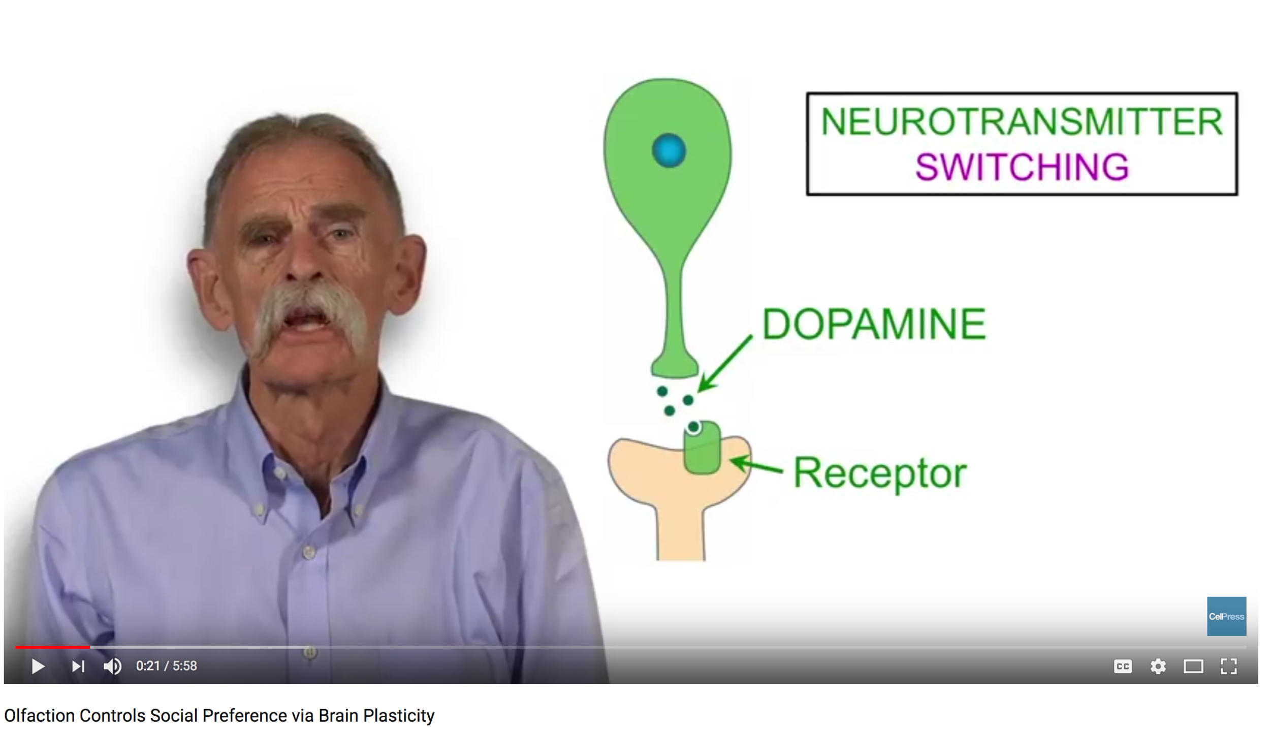
Olfaction Controls Social Preference via Brain Plasticity


Video abstract for our Neuron paper. DOI: https://doi.org/10.1016/j.neuron.2017.08.023

For more information, see Dulcis et al., Neuron 95/6, http://www.cell.com/neuron/fulltext/S0896-6273(17)30738-9. Acquisition of social preference for relatives versus strangers is dynamic, but the developmental regulatory mechanisms have been unclear. Dulcis et al. found that sustained exposure to olfactory cues converts non-kin aversion to attraction via neurotransmitter switching coordinated by microRNAs.

Mnemonic microRNAs help make memories

From: In Focus (Sep 19, 2011)

Researchers find that two microRNAs prime the brain to learn. Learning and memory depend on the brain's plasticity, its ability to rewire itself. Lippi et al. reveal that two microRNAs (miRNAs) help the brain maintain this flexibility by dismantling unneeded synapses between neurons (1).

F1.medium.gif

FOCAL POINT Kenneth Young (left), Giordano Lippi (center), and colleagues (not pictured) tracked down two miRNAs that induce synaptic plasticity in the brain. A control hippocampal dendrite (top right) possesses filopodia and several kinds of spines, including mushroom-shaped spines. But a dendrite that overproduces miR-29a (bottom right) displays fewer mushroom-shaped spines and more spindly filopodia.

When a rat memorizes the route through a maze or a singer learns the lyrics to a song, the neural changes in their brains typically include growth or modification of dendritic spines, the antennae that receive impulses from axons of neighboring neurons. Accumulating evidence suggests that miRNAs have a big influence on dendritic spines. They can, for instance, fine-tune protein production within a spine (2). Some research also indicates that miRNAs control the formation, structure, and function of dendritic spines (3, 4). However, researchers don't fully understand how miRNAs affect neural plasticity.

Lippi et al. wanted to identify miRNAs that might shape synapses, so they dosed mice with nicotine, cocaine, or amphetamine, all of which promote brain rewiring. The team then analyzed different brain regions to pinpoint miRNAs whose levels rose after treatment with the drugs. Quantities of 32 miRNAs climbed in most brain regions, the researchers found. To ascertain whether these miRNAs promote brain plasticity, the researchers measured their levels in cultured neurons that were just beginning to synapse with each other. They detected surges in two pairs of related miRNAs: miR-29a/miR-29b and miR-182/miR-183. Lippi et al. also found that increased firing by neurons boosted the levels of these miRNAs, suggesting that synaptic activity regulates the expression of the miRNAs.

But are the miRNAs molding synaptic architecture? To find out, the researchers engineered cultured hippocampal neurons to overproduce each of the four miRNAs. Dendritic spines start out as simple protrusions known as filopodia, and they often mature into more permanent, mushroom-shaped structures. In control neurons, mushroom-shaped dendritic spines predominated, the scientists found. But neurons that carried extra miR-29a or miR-29b sported fewer mushroom-shaped spines and more filopodia. By contrast, miR-182 and miR-183 had little effect on dendritic spines. These results suggest that miR-29a and miR-29b regulate the formation and maintenance of synapses, returning them to a less specialized state.

We think that these miRNAs maintain plasticity in neurons

The researchers uncovered the mechanism. Using RNAi, Lippi et al. depleted nine likely targets of the miRNAs from cultured neurons. Knocking down one of the proteins, Arpc3, produced the same effects on dendritic spines as boosting levels of miR-29a and miR-29b. Arpc3 is part of the ARP2/3 actin-nucleation complex, which spurs actin to branch, a key step in the maturation of dendritic spines. Further experiments showed that miR-29a and miR-29b slash Arpc3 levels.

Lippi et al. also found evidence that the miRNAs promote learning in vivo. The team measured miRNA levels in mice that had undergone fear conditioning, in which the animals learn to associate a tone with electric shocks to their feet. Amounts of miR-29a and miR-29b in the hippocampus shot up after the training, and levels of Arpc3 fell.

Instead of building new connections between neurons, miR-29a and miR-29b appear to thwart them, preventing dendritic spines from maturing by blocking the ARP2/3 complex that spurs actin polymerization. “We think that these miRNAs maintain plasticity in neurons,” says first author Giordano Lippi. The molecules “could be part of a mechanism by which spines are eliminated when they aren't necessary anymore,” he says. By pruning nonessential spines, the miRNAs might ensure that neurons can extend new spines when they are required.

How important these miRNAs are for editing neural connections in the brain remains to be seen. Drug addiction can lead to construction of new synapses, so one question is whether drugs inhibit these miRNAs to allow neural connections to form and consolidate. The miRNAs could also be important in conditions in which memory and learning falter, such as Alzheimer's disease.


Shaping Early Networks to Rule Mature Circuits: Little MiRs Go a Long Way

From: Neuron Preview (Dec 21, 2016). Authors: Andre Marques-Smith, Emilia Favuzzi, and Beatriz Rico

Normative cortical processing depends on precise interactions between excitatory and inhibitory neurons. In this issue of Neuron, Lippi et al. (2016) identify miR-101 as a master regulator coordinating molecular programs during development that ultimately impact the activity of mature networks.

Main Text

Neural computation relies on the precise organization of synaptic connections among different neuronal subtypes. Interactions between excitatory pyramidal neurons and inhibitory GABAergic interneurons are particularly important, as neuronal circuits can only operate effectively within certain bounds of excitation and inhibition (Isaacson and Scanziani, 2011). This is critical not only for the information processing that supports animal behavior but also because overstepping these boundaries can lead to neurodevelopmental and neurological disorders, including autism, schizophrenia, and epilepsy (Paz and Huguenard, 2015, Marín, 2016).

During brain development a plethora of turbulent events will frame mature neural circuits: endogenous spontaneous rhythms give way to sensory-driven activity, GABA switches polarity, canonical circuits are formed, potentiated, and refined, and eventually synapses elevate their threshold for plasticity, narrowing integration windows to become fast, precise reporters of spiking activity. Each of these processes is regulated by dynamic programs of gene expression, which are tuned by neural activity in a bidirectional manner. What could quickly become a neural cacophony actually plays out as a beautifully orchestrated symphony; transcriptional programs regulate expression of ion channels, neurotransmitter receptors, and transporters, restraining patterns of network activity and controlling the transition between them. The intimate association of several such developmental processes—e.g., dendritic arbor elaboration and synapse formation—and the need to concertedly switch transcription on or off for different genes requires centralized regulation of gene cohorts to effect on-going neural genetic programs. MicroRNAs (miRs) are small non-coding RNAs that function as post-transcriptional regulators of gene expression holding the ability to simultaneously regulate multiple genes in the context of complex regulatory networks (McNeill and Van Vactor, 2012). miRs provide mechanisms of regulation that are fast, flexible, and reversible and as such, well-suited for the complexities of neural circuit wiring. They appear thus as ideal candidates to tightly regulate and tune developmental gene programs during the assembly of neuronal circuits. In a series of elegant experiments, Lippi et al. (2016) discover that microRNA 101 (miR-101) synergistically regulates expression of several genes for the common goal of constraining excitation in hippocampal circuits.

Lippi et al. (2016) carried out a thorough screening to identify sequenced miRs in the developing hippocampus at postnatal day 12 (P12), a critical developmental window, curating a list of candidates well suited for neural developmental processes. Then, based on: (1) abundance, (2) upregulation during development, (3) enrichment in Argonaute complexes (Ago, effector of miR function), and (4) published targets of miRs involved in neural differentiation, they identified miR-101. Transient (P2–P9) and localized inhibition of miR-101 resulted in a lasting adult phenotype characterized overall by hyper-excitability. Adult hippocampal pyramidal neurons displayed increased firing rates in vivo, as well as elevated frequency and amplitude of spontaneous excitatory post-synaptic currents (sEPSCs) in vitro. Calcium imaging experiments revealed higher proportions of active neurons at any one time, as well as an overall increase in frequency of calcium (putative spiking) events. By using behavioral tests that depend on hippocampal function, Lippi et al. (2016) showed that blocking miR-101 in early postnatal life—but not in adult—led to lasting deficits in context-dependent associative memory, spatial working memory, and spatial episodic-like memory. These findings are particularly relevant for neurodevelopmental disorders, as they link the transient early inhibition of miR-101 to impaired cognitive function in the adult.

To identify the mechanism by which miR-101 regulates the establishment of a balanced network, they searched for miR-101 targets. Using a combination of in vitro and in vivo approaches, Lippi et al. (2016) revealed several candidates, including the sodium-potassium-chloride co-transporter 1 (NKCC1). Across multiple brain regions, downregulation of this chloride importer and upregulation of the chloride exporter KCC2 underlies the developmental shift in chloride reversal potential and consequent maturation of GABAergic signaling from depolarizing to hyperpolarizing (Ben-Ari, 2002). Indeed, blocking miR-101 in vivo resulted in increased NKCC1 expression by release from miR-101 repression and a relatively depolarized EGABA at P8. In contrast, KCC2 expression was unchanged, suggesting that a distinct developmental genetic program regulates KCC2 levels. By disrupting miR-101-NKCC1 interaction without affecting other miR-101 targets, Lippi et al. (2016) elegantly demonstrate that miR-101 regulation of NKCC1 mRNA alone was responsible for the delayed maturation of the GABA reversal potential. Giant depolarizing potentials (GDPs) synchronize activity and promote synaptic plasticity between pyramidal neurons (Allène et al., 2008). Furthermore, early GABAergic activity is required for dendritic elaboration (Cancedda et al., 2007). Therefore, a sustained depolarizing action of GABA in miR-101 blocking experiments could affect both synaptic stabilization and dendritic development leading to an exuberant excitatory network. However, specific de-repression of NKCC1 without affecting other miR-101 in vivo only explained the increased rate of synchronous calcium events and a modest elevation in miniature EPSC frequency in vitro, causing no discernible effect on overall rate of calcium, proportion of active ensembles, or double synchronized events, hallmark features of the miR-101 phenotype.

Given the partial phenotype of prolonged NKCC1 expression, Lippi et al. (2016) hypothesized that the effect of miR-101 inhibition was achieved through multi-level targeting of several genes within a biological network. They explored this possibility by combining the top targets for miR-101 into groups, according to their known developmental ontology effects (“Pre-synaptic,” “Glial,” and “Excitability”). In addition to NKCC1, the “Pre-synaptic” group included two genes involved in the formation and stabilization of presynaptic inputs, Ank2 and Kif1a. Lippi et al. (2016) elegantly dissected the contribution of these genes, finding that NKCC1 targeting by miR-101 limits dendritic length while complementary repression of Kif1a and Ank2 is required to restrict excitatory synaptic density. As a result, continued expression of NKCC1 and the genes in the “Pre-synaptic” group mimicked the increased levels of activity, mainly because of the occurrence of more asynchronous calcium events. Next, de-repression of NKCC1 and two genes, the cholesterol transporter Abca1 and the hydrolase Ndrg2 (“Glial” group), enriched in glial cells with a role in neurite growth, was responsible for the increase in the size of cell ensembles recruited in each synchronous event. Releasing the expression of genes involved in regulating neuronal excitability (“Excitability” group), along with NKCC1, increased the number of double events. Thus, each group of genes accounted for unique aspects of the multi-tier regulatory control of miR-101 and together they dictate the precise code for a balanced development of neural circuits (Figure 1).

Although previous studies have proposed that miRs function in shaping the neuronal landscape (see McNeill and Van Vactor, 2012), the work of Lippi et al. (2016) constitutes the first demonstration that simultaneous regulation of multiple target genes by a single miR during a critical developmental window orchestrates convergent molecular programs that ultimately sculpt a stable mature neuronal network (Figure 1). Also, it is important to emphasize that this study has been carried out using in vivo models where the cellular context is intact, demonstrating a more physiological function of the miR. In sum, Lippi et al. (2016) reveal here a set of interesting results with implications not only for miR biology and function, but also for the regulation of excitatory-inhibitory balance and neurodevelopmental processes.

Figure 1. 

miR-101 Regulates the Development of Neural Circuits Shaping Mature Networks in Adult. 

It remains unknown why the long-lasting effects caused by early transient miR-101 blockage were not compensated homeostatically. It is well documented that neurons and networks are highly reactive to, and capable of compensating for, changes in their excitatory-inhibitory environment (Xue et al., 2014). It is surprising therefore that the hippocampal network did not respond to unfettered excitation through release from miR-101 regulation by increasing inhibition. Indeed, increases in excitation occurred in the absence of proportional changes in inhibitory currents, suggesting the presence of exuberant excitatory circuits rather than dis-inhibition. This is particularly intriguing since miR-101 is also expressed in interneurons. Interestingly, the lack of epileptiform activity in such an excitable network in itself suggests that subtler forms of compensation occurred and went undetected, preventing the emergence of pathology. An attractive possibility is that miR-101 itself regulates inhibitory synapse formation while it constrains excitation, and blockade of its action prevented emergence of an inhibitory compensatory response. It will be interesting to determine the role of miR-101 in different types of GABAergic cells. Inhibition synchronizes and sharpens excitatory responses in many brain areas, and its impairment could increase “noise” in learning and cognition, partially accounting for some of the observed cognitive effects of miR-101 inhibition described by Lippi et al. (2016).

What determines the changes in the expression of miR-101 in the first place? Is it the result of specific activity patterns or is it intrinsically determined? Pyramidal neurons receive inhibition in proportion to their afferent synaptic excitation levels, meaning E/I balances across cells are stable even though afferent excitation levels differ widely (Xue et al., 2014). How does the genetic regulation of E/I balance, through miR-101 and other actors, operate at the individual cell level? The simplest hypothesis is that genes regulating E/I balance are responsive to neural activity. miRs have indeed been previously linked to activity (McNeill and Van Vactor, 2012). Could miR-101, for instance, sense chronic increases in excitatory activity and increase repression of its downstream targets?

Additional work will be needed to examine whether miR-101 plays a similar role in other brain regions such as the neocortex. This will help to determine whether the regulatory developmental program described by Lippi et al. (2016) represents a general mechanism to constrain excitation in the brain. This is particularly relevant since neural circuits show exquisite fine structure, with spatially proximal cells often participating in completely different microcircuits and subnetworks (Lee et al., 2014). These channels of information may not have the same ratio of excitation and inhibition and may differentially impact neural function. Could miRs help sculpt an additional level of circuit-specificity, beyond cell-type rules of innervation? Some data in Lippi et al. (2016)’s work hints at pathway-specific regulation, e.g., discrepant effect of NKCC1 de-repression on the secondary branches in CA1 and in CA3 or over-representation of mossy fiber input. It would be of great interest to extend these observations with pathway and cell-type-specific methods.

Because of its ability to regulate multiple key aspects of brain development, it is not surprising that miR-101 has a role in many neuropsychiatric disorders (Lippi et al., 2016, Figure 1). Interestingly, the prevalent view in the field is that although development is a continuous process, there are specific sensitive windows—“critical periods”—in which modifications in network organization have long-lasting impact over the lifespan (Marín, 2016). These sensitive periods are pivotal milestones for the assembly of excitatory and inhibitory circuits. Therefore, understanding how the relative bounds of excitation and inhibition are developmentally established, maintained, and shifted is an exciting topic of research, increasingly attracting interest in Neuroscience. Indeed, unveiling the main regulators of these processes might be key for early interventions to restore normal brain function (Marín, 2016). Future work uncovering the function of miRs in neural circuit development promises to shed light on potential therapeutic targets for neurodevelopmental disorders.

Acknowledgments

Work in B.R. laboratory is supported by the European Research Council (ERC-2012-StG 310021). B.R. is Wellcome Trust Investigator.

Scents and Social Preference: Neuroscientists ID the Roots of Attraction

From: UC San Diego News Center (Aug 2, 2017)

A baby lamb is separated from its family. Somehow, in vast herds of sheep that look virtually identical, the lost youngling locates its kin. Salmon swim out to the vast expanses of the sea and migrate back home to their precise spawning grounds with bewildering accuracy.

Scientists have long known about such animal kinship attachments, some known as “imprinting,” but the mechanisms underlying them have been hidden in a black box at the cellular and molecular levels. Now, biologists at the University of California San Diego have unlocked key elements of these mysteries, with implications for understanding social attraction and aversion in a range of animals and humans.

Davide Dulcis of UC San Diego’s Psychiatry Department at the School of Medicine, Giordano Lippi, Darwin Berg and Nick Spitzer of the Division of Biological Sciences and their colleagues published their results in the August 31, 2017 online issue of the journal Neuron.

dulcis-neuron-450.jpg

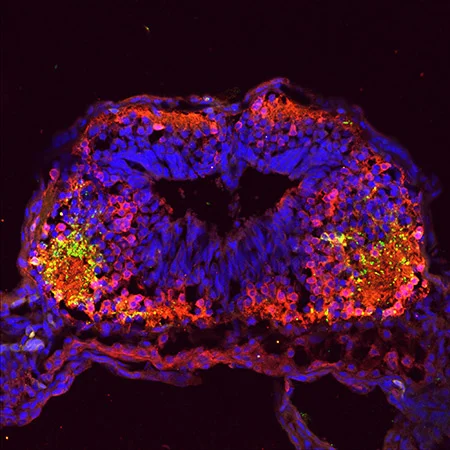
Figure 1.

A confocal image of a tadpole brain reveals dopaminergic (green) neurons, the type increased in typical kinship recognition, and GABAergic (red) neurons, those elevated in cases of expanded social kinship.

In a series of neurobiological studies stretching back eight years, the researchers examined larval frogs (tadpoles), which are known to swim with family members in clusters. Focusing the studies on familial olfactory cues, or kinship odors, the researchers identified the mechanisms by which two- to four-day-old tadpoles chose to swim with family members over non-family members. Their tests also revealed that tadpoles that were exposed to early formative odors of those outside of their family cluster were also inclined to swim with the group that generated the smell, expanding their social preference beyond their own true kin.

The researchers discovered that this change is rooted in a process known as “neurotransmitter switching,” an area of brain research pioneered by Spitzer and further investigated by Dulcis in the context of psychostimulants and the diseased brain. The dopamine neurotransmitter was found in high levels during normal family kinship bonding, but switched to the GABA neurotransmitter in the case of artificial odor kinship, or “non-kin” attraction.

“In the reversed conditions there is a clear sign of neurotransmitter switching, so now we can see that these neurotransmitters are really controlling a specific behavior,” said Dulcis, an associate professor. “You can imagine how important this is for social preference and behavior. We have innate responses in relationships, falling in love and deciding whether we like someone. We use a variety of cues and these odorants can be part of the social preference equation.”

The scientists took the study to a deeper level, seeking to find how this mechanism unfolds at the genetic level.

Sequencing helped isolate two key microRNAs, molecules involved in coordinating gene expression. Sifting through hundreds of possibilities, they identified microRNA-375 and microRNA-200b as the key regulators mediating the neurotransmitter switching for attraction and aversion, affecting the expression of genes known as Pax6 and Bcl11b that ultimately control the tadpole’s swimming behavior.

“MicroRNAs were ideal candidates for the job,” said Lippi, a project scientist in Berg’s laboratory in the Division’s Neurobiology Section. “They are post-transcriptional repressors and can target hundreds of different mRNAs to consolidate specific genetic programs and trigger developmental switches.”

The study began in 2009 and deepened in size and scope over the years. Reviewers of the paper were impressed with the project’s breadth, including one who commended the authors “for this heroic study which is both fascinating and comprehensive.”

“Social interaction, whether it’s with people in the workplace or with family and friends, has many determinants,” said Spitzer, a distinguished professor in the Division of Biological Sciences, the Atkinson Family Chair and co-director of the Kavli Institute for Brain and Mind at UC San Diego. “As human beings we are complicated and we have multiple mechanisms to achieve social bonding, but it seems likely that this mechanism for switching social preference in response to olfactory stimuli contributes to some extent.”

In addition to Dulcis, Lippi, Berg and Spitzer, coauthors of the paper include Christiana Stark and Long Do.

The National Institutes of Health-National Institute of Neurological Disorders and Stroke supported the research (R01 NS015918 awarded to Spitzer and R01NS057690 awarded to Dulcis and Spitzer).